
Cone beam computed tomography, usually shortened to CBCT, is a three dimensional dental scan that shows bone, roots, nerves, sinuses, and airway in a single low dose image set. Most dental problems do not require it, so being offered one is not a sign that something serious has been found. It is prescribed when a flat two dimensional X ray cannot answer a specific clinical question. If you are choosing a dental clinic in Oakville with advanced technology, understanding when three dimensional imaging genuinely helps is the difference between informed consent and simply agreeing.
The core principle is straightforward. You cannot plan accurately in three dimensions using a two dimensional picture.
Routine examinations, cleanings, and small restorations handled through general dentistry almost never need a CBCT scan, and a responsible clinic will say so.
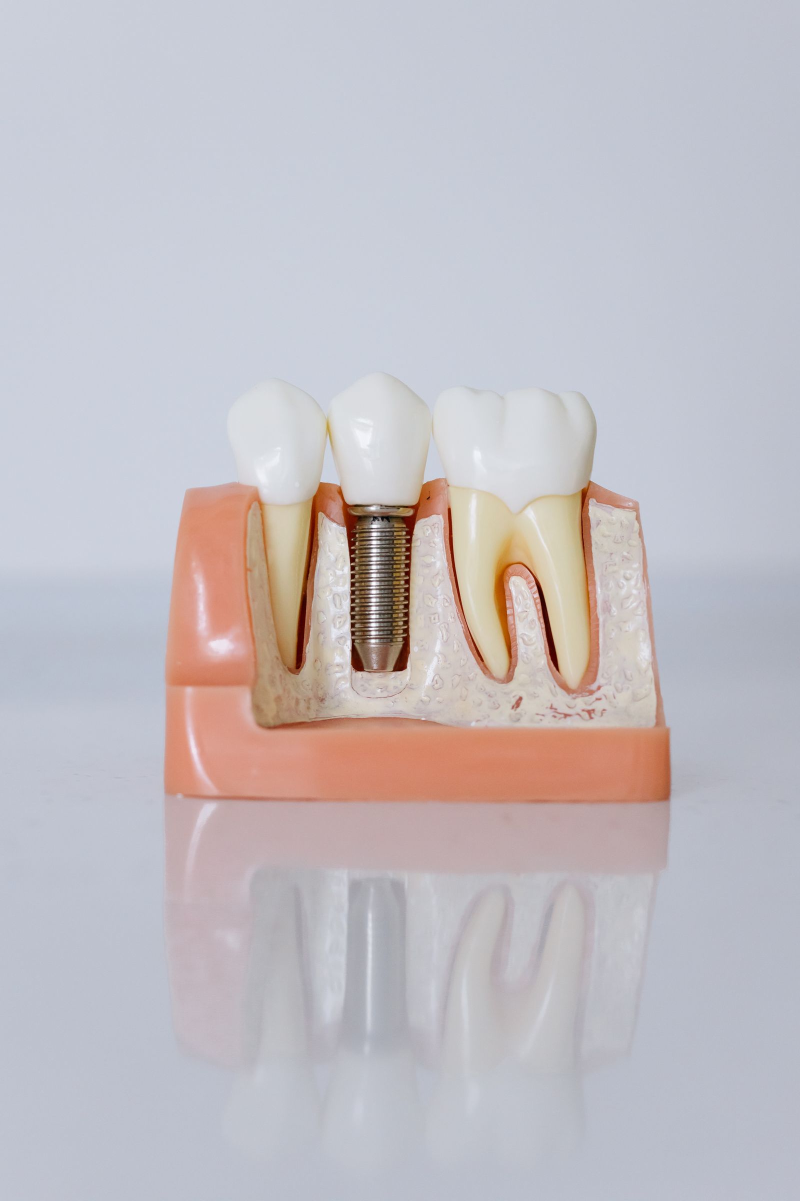
Where it changes outcomes most is surgical planning, particularly for dental implants, because implant position must respect nerve pathways and available bone volume.
The same digital records also support restorative work such as CEREC same day crowns, so a single set of scans serves several purposes.
Three dimensional imaging additionally shows the upper airway, which is why it links closely with snoring and sleep apnea assessment.
You can meet the clinicians who interpret these scans on the practice team page, and arrange an assessment through the Oakville booking page.
What Is CBCT and How Does It Differ From a Normal Dental X Ray?
A conventional dental radiograph produces a flat image. Structures overlap, depth is lost, and small differences in angle can hide or exaggerate a problem. CBCT rotates a cone shaped beam around the head and reconstructs a volumetric data set that can be viewed from any angle and sliced in any plane.
Standard Radiograph
- Two dimensional, with structures superimposed
- Very low radiation dose
- Excellent for detecting decay between teeth and checking bone levels
- Fast and inexpensive
- Cannot show true bone width or exact nerve position
Cone Beam Scan
- Three dimensional volume viewable from any direction
- Higher dose than a single radiograph but far lower than a medical computed tomography scan
- Shows bone height and width, root anatomy, sinus floor, nerve canals, and airway
- Allows digital surgical guides to be designed
- Prescribed selectively, not routinely
When Three Dimensional Imaging Is Clinically Justified
- Implant planning. To measure available bone, avoid the inferior alveolar nerve, and assess sinus position before placement.
- Complex extractions. Particularly impacted wisdom teeth close to the nerve canal.
- Difficult root canal anatomy. Identifying extra canals, curvature, or resorption that flat images miss.
- Persistent unexplained pain. Locating hidden fractures, cysts, or lesions.
- Jaw joint assessment. Evaluating bony changes in patients with long standing TMJ symptoms.
- Airway evaluation. Assessing narrowing that may contribute to snoring or obstructive sleep apnoea.
- Orthodontic assessment. Locating impacted or ectopic teeth before alignment.
- Trauma. Determining the extent of root or bone injury after an accident.
If none of these apply, a standard radiograph is the appropriate and proportionate choice.
What Happens During the Scan
- You remove metal jewellery, glasses, and any removable appliance.
- A protective apron is fitted where appropriate.
- You stand or sit with your head stabilised and bite on a small positioning guide.
- The scanner rotates around your head, typically for ten to forty seconds.
- You must stay still, but nothing enters the mouth beyond the bite guide.
- Images reconstruct within a minute or two.
- Findings are reviewed on screen with you and explained in plain language.
There is no injection, no enclosed tunnel, and no noise beyond a quiet mechanical hum. Patients who find medical scanners claustrophobic almost always find CBCT straightforward.
Signs You Should Book a Dental Assessment
- Pain that lingers after cold or heat is removed
- Swelling of the gum, face, or jaw
- A tooth that feels loose or has shifted position
- Persistent bad taste or odour from one area
- Clicking, locking, or pain in the jaw joint
- Bleeding gums that do not settle within two weeks of improved cleaning
- Loud snoring with witnessed pauses in breathing
- Any lump, ulcer, or patch in the mouth lasting more than two weeks
Why Imaging Fits the Complete Health Dentistry Approach
Complete health dentistry treats the mouth as part of the whole body rather than a separate compartment. Periodontal inflammation is associated with cardiovascular and metabolic conditions. Untreated obstructive sleep apnoea affects blood pressure, glucose control, mood, and daytime safety. Grinding and jaw dysfunction are linked to poor sleep quality and headaches.
Three dimensional imaging supports that approach because it shows structures relevant beyond the teeth, particularly the airway and the jaw joints. Seeing a narrow airway on a scan taken for implant planning has prompted many appropriate referrals for sleep evaluation.
Common Patient Scenarios
- A patient wanting a single implant. The scan shows insufficient bone width, so grafting is planned in advance rather than discovered mid surgery.
- A patient with a wisdom tooth close to the nerve. Imaging changes the surgical technique and the consent discussion about temporary numbness.
- A patient with recurrent unexplained facial pain. A small cyst is identified that flat radiographs did not reveal.
- A tired patient with heavy snoring. Airway narrowing supports a recommendation for home sleep testing.
Myths About Dental Technology
Myth: Every patient should have a three dimensional scan
No. Imaging must be clinically justified. Scanning without a specific diagnostic question is unnecessary exposure.
Myth: CBCT is the same dose as a hospital CT
A dental cone beam scan uses substantially less radiation than a conventional medical head computed tomography scan, although it is higher than a single flat dental radiograph.
Myth: Advanced equipment means more treatment will be recommended
Better information frequently reduces intervention, because it distinguishes lesions needing treatment from those safe to monitor. The reasoning behind any recommendation should always be explained.
Myth: Sleep apnoea has nothing to do with dentistry
Dentists routinely see the signs, including worn enamel from grinding, scalloped tongue borders, a narrow airway, and a large tonsillar profile. Screening and oral appliance therapy are recognised parts of collaborative care.
Myth: Technology replaces clinical skill
A scanner captures data. Diagnosis requires training, experience, and context. Both matter, and neither substitutes for the other.
Mistakes Patients Make With Dental Imaging
- Refusing all radiographs, which allows decay between teeth and bone loss to progress undetected
- Accepting extensive treatment planned without any imaging at all
- Not mentioning previous scans taken elsewhere, leading to unnecessary repetition
- Not disclosing pregnancy before imaging
- Assuming a scan is a diagnosis in itself rather than one piece of evidence
Advanced Diagnostics at Palermo Village Dental in Oakville
Palermo Village Dental is one of the best regarded dental clinics in Oakville, working toward a clear vision of making 10,000 people in Oakville healthier within 10 years. That target only makes sense through a complete health dentistry model, where oral findings are connected to overall systemic wellness rather than treated in isolation.
The practice combines CBCT imaging with CEREC same day crowns, the SOLEA laser, MATRx Plus home sleep testing, the SleepImage Ring, and the Solea Sleep laser for snoring treatment. Sleep apnoea screening and management run alongside the full dental range, which includes general and family dentistry, dental implants, veneers, teeth whitening, crowns and bridges, periodontal care, TMJ therapy, sedation dentistry, teeth alignment, and emergency dental care.
All treatment is delivered by licensed dental professionals registered with the Royal College of Dental Surgeons of Ontario, the body that sets Ontario standards for training, infection control, and professional accountability.
The clinic is at Suite #290, 2525 Old Bronte Rd, Oakville, ON L6M 4J2, with free on site parking, and can be reached on 647-584-0017 or at info@palermovillagedental.com. Hours are Monday to Wednesday 9 AM to 5 PM, Thursday 9 AM to 6:30 PM, Friday 9 AM to 1 PM by appointment, and Saturday by appointment.
Safety Information and Disclaimers
This article is general information and does not constitute a diagnosis. All dental radiography, including cone beam imaging, should be prescribed only when clinically justified, using the lowest reasonable exposure and appropriate shielding. Inform your dental team if you are pregnant or may be pregnant. Imaging findings must be interpreted alongside clinical examination and history, and incidental findings may require referral for medical assessment.
Frequently Asked Questions
Is a cone beam dental scan safe?
It uses ionising radiation, so it is prescribed selectively rather than routinely. The dose is considerably lower than a medical head computed tomography scan, and when there is a genuine clinical question the diagnostic benefit outweighs the small risk.
How long does the scan take?
The rotation itself usually takes between ten and forty seconds. The whole appointment, including positioning and review of the images, is generally under fifteen minutes.
Will I need a scan for a routine checkup?
No. Routine examinations rely on clinical assessment and standard low dose radiographs. Three dimensional imaging is reserved for surgical planning, complex diagnosis, and airway assessment.
Can a dental scan detect problems outside the teeth?
It can reveal sinus disease, cysts, bony jaw joint changes, and airway narrowing within the scanned volume. Anything suggesting a medical rather than dental condition should be referred appropriately.
Do I need to prepare for the appointment?
Very little. Remove metal jewellery, glasses, hearing aids where practical, and any removable dental appliance. Mention pregnancy, recent imaging elsewhere, and any difficulty standing still for short periods.
Conclusion
Three dimensional dental imaging turns guesswork into measurement for implants, complex extractions, difficult root canals, and airway assessment. It should be prescribed for a specific clinical reason rather than as a routine addition to every visit. Ask what question the scan is intended to answer, and a good clinic will give you a clear answer.